
Keywords: Alzheimer’s Disease, APOE ε4, Glucosamine, Hexosamine Biosynthetic Pathway (HBP), Hyperglycosylation, Mild Cognitive Impairment, O-GlcNAcylation, Tau Protein
Introduction
Alzheimer’s disease (AD) is the most prevalent form of dementia, currently affecting an estimated 55 million people worldwide, with projections exceeding 150 million by 2050 as populations age. Despite decades of research and the recent approval of amyloid-targeting therapies, the disease remains incurable, and its full pathogenic complexity is yet to be mapped. The canonical hallmarks of AD, amyloid-beta (Aβ) plaques and neurofibrillary tangles (NFTs) composed of hyperphosphorylated tau have dominated the research landscape. However, an expanding body of evidence now implicates upstream metabolic dysregulation as a critical and potentially modifiable contributor to disease initiation and progression [2].
Among the many post-translational protein modifications studied in the context of neurodegeneration, glycosylation, the enzymatic attachment of sugar molecules to proteins has emerged as particularly significant. Brain glycosylation functions physiologically to direct protein folding, regulate receptor signaling, modulate synaptic plasticity, and maintain cellular homeostasis. However, when this system becomes dysregulated, the consequences for neuronal integrity are profound [3,4].
A landmark study published in Nature Metabolism in June 2026 by Hawkinson and colleagues introduced a compelling new framework: hyperglycosylation, the pathological excess of sugar-chain addition to brain proteins as a bona fide metabolic driver of AD. Crucially, this research identified a common over-the-counter supplement, glucosamine, as a modifiable factor capable of exacerbating brain hyperglycosylation and accelerating disease progression. This finding carries significant public health implications, given that glucosamine is taken regularly by an estimated one-fifth of adults in the United States, Australia, and the United Kingdom for the management of osteoarthritis-related joint pain [1,5].
This review article examines the biological basis of hyperglycosylation in AD, appraises the translational evidence from animal models and human cohort studies, contextualizes the risk associated with glucosamine supplementation, particularly for those carrying the APOEε4 allele and discusses emerging therapeutic and preventive strategies relevant to longevity and metabolic medicine.
Protein Glycosylation in The Brain: Physiology and Pathological Dysregulation
Glycosylation is one of the most common post-translational modifications (PTMs) in eukaryotic cells, affecting an estimated 50-70% of all human proteins. In the central nervous system (CNS), two principal forms are relevant to AD: N-linked glycosylation, which occurs on asparagine residues within the endoplasmic reticulum (ER) as proteins are being folded, and O-linked N-acetylglucosaminylation (O-GlcNAcylation), a cytoplasmic and nuclear modification that dynamically cycles on serine and threonine residues of intracellular proteins [4].
O-GlcNAcylation is particularly important in the neuroscience of AD because it shares regulatory sites with phosphorylation on tau protein. Physiological levels of O-GlcNAc on tau promote microtubule stability and reduce pathological tau phosphorylation. This reciprocal relationship, sometimes termed the ‘yin-yang’ hypothesis, suggests that a balanced O-GlcNAc state is neuroprotective, while both excessive and deficient O-GlcNAcylation can disturb downstream signaling [6,7].
The substrate for O-GlcNAcylation is UDP-GlcNAc, synthesized via the hexosamine biosynthetic pathway (HBP). The HBP acts as a metabolic sensor: it integrates glucose, glutamine, acetyl-CoA, and uridine-5′-triphosphate (UTP) to produce UDP-GlcNAc, the sugar donor used by O-GlcNAc transferase (OGT). Accordingly, elevated glucose flux, as seen in hyperglycaemia, insulin resistance, or metabolic syndrome drives upregulation of the HBP and may secondarily increase glycosylation in the brain [4,8].
Under physiological conditions, this process is tightly regulated. O-GlcNAcase (OGA) removes the GlcNAc modification, and the cycle of addition and removal is balanced. In the AD brain, however, this equilibrium is disrupted: a state of hyperglycosylation emerges, characterized by excessive N-glycan chains distributed aberrantly across cortical, hippocampal, and thalamic neurons, precisely the region’s most vulnerable to AD pathology [1].
Hyperglycosylation as a Hallmark of Alzheimer’s Disease: Human Evidence
The study by Hawkinson et al. employed an integrative multiomics approach combining spatial metabolomics, lipidomics, and glycomics to examine brain tissue from both transgenic AD mouse models and post-mortem human samples. Human frontal cortex specimens from AD patients and age, sex-, and post-mortem interval-matched controls were analyzed using matrix-assisted laser desorption/ionization (MALDI) imaging mass spectrometry, enabling spatially resolved quantification of glycan levels across grey and white matter [1].
The results were striking. Glycosylation was elevated 10- to 20-fold in AD brain regions compared to controls, particularly in the cortex, hippocampus, and thalamus, structures central to memory encoding and retrieval, and well-documented sites of early AD pathology. This pattern was independently validated in a second human cohort, confirming its reproducibility. Spatially, the hyperglycosylation signal was most intense in grey matter regions harboring neuritic plaques and NFTs, suggesting a mechanistic relationship with established pathological substrates [1].
Advanced spatial isotopic tracing using pulse-chase analysis of N-linked glycans demonstrated that this hyperglycosylation phenotype is driven primarily by increased glycan biosynthesis rather than impaired glycan clearance. This distinction is clinically important: it implies that interventions targeting synthetic pathways upstream, such as inhibiting glutamine: fructose-6-phosphate transaminase (GFPT), the rate-limiting enzyme of the HBP, may be therapeutically viable [1].
The Braak Staging Correlation: Glycosylation as a Progressive Disease Marker
The Braak and Braak neuropathological staging system, first described in 1995, provides a six-stage framework for classifying the topographic progression of NFTs from the entorhinal cortex (stages I-II) through limbic structures (stages III-IV) to isocortical regions (stages V-VI). This hierarchical staging system correlates closely with the clinical severity of dementia and has been validated using in vivo tau positron emission tomography (PET) imaging [9,10].
Hawkinson et al. found that brain hyperglycosylation levels tracked linearly and significantly with Braak stages across the full disease spectrum. At Braak stage 0, representing essentially normal brain tissue, glycosylation was at its nadir. Levels rose progressively at stages I-II, III-IV, and peaked at stages V-VI, corresponding to severe cortical NFT burden and profound clinical dementia. This dose-response relationship is hallmarking epidemiological evidence for a mechanistic link rather than mere epiphenomenon: the more advanced the disease, the more pronounced the hyperglycosylation [1].
This finding aligns with broader evidence from metabolic profiling studies demonstrating region-specific metabolic derangements that intensify with AD severity. Crucially, the distribution of hyperglycosylation was not uniform but concentrated in anatomical regions known to be most severely affected by NFT pathology, supporting the hypothesis that glycan dysregulation is integral to, rather than a consequence of progressive neurodegeneration [11].
From Correlation to Causation: Experimental Evidence in Preclinical Models
Demonstrating causality requires interventional evidence: blocking a proposed mechanism should protect against the disease phenotype, while enhancing it should worsen outcomes. Hawkinson et al. conducted precisely such experiments using two well-validated transgenic mouse models of AD: the 5xFAD model, which overexpresses five human familial AD mutations in amyloid precursor protein (APP) and presenilin-1 (PSEN1) and manifests Aβ accumulation and cognitive decline from 4-6 months of age; and the PS19 model, which expresses a P301S mutation in human tau and develops robust NFT pathology [1,12].
Genetic knockdown of glycan biosynthetic enzymes, specifically those catalyzing N-glycan chain elongation in both 5xFAD and PS19 mice resulted in significant improvements in spatial and working memory compared to unmodified AD controls. Complementary pharmacological inhibition using small-molecule glycan biosynthesis inhibitors replicated this benefit. These findings establish that excessive glycosylation is not merely a correlate of AD pathology but actively contributes to neuronal dysfunction and cognitive impairment [1].
Mechanistically, hyperglycosylation is proposed to disrupt protein-protein interactions critical to synaptic transmission, impair proteasomal and lysosomal clearance of misfolded proteins, and promote the aggregation of tau into insoluble NFTs by competing with protective O-GlcNAc modifications at key phosphorylation sites. The net effect is a self-reinforcing cycle: as glycan burden increases, neuronal proteostasis deteriorates, Aβ and tau accumulate, and cognitive deficits deepen [6,7,8].
Glucosamine Supplementation: a Common Supplement with Uncommon Neurological Risk
Pharmacology and Prevalence
Glucosamine is an endogenous amino sugar and structural precursor of glycosaminoglycans and proteoglycans, which are essential components of articular cartilage. As an oral supplement, typically administered at 1,500-2,500 mg daily either as glucosamine sulphate or glucosamine hydrochloride, it has been marketed extensively for the relief of osteoarthritis symptoms. Its use is widespread: surveys indicate regular use among approximately 20% of adults in the United States, the United Kingdom, and Australia, corresponding to tens of millions of individuals [5,13].
Until recently, the safety profile of glucosamine was considered broadly favorable, with principal concerns limited to mild gastrointestinal side effects, potential blood glucose perturbations in diabetic patients, and possible cardiovascular risks in susceptible populations. The notion that it might influence central nervous system pathology was largely unexplored [13,14].
Brain Penetration and Glycan Incorporation
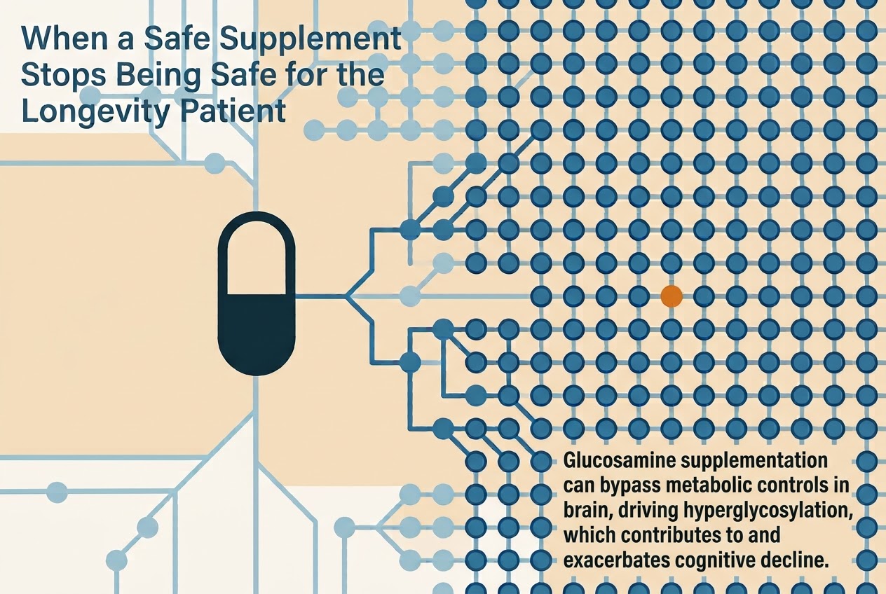
Hawkinson et al. demonstrated that orally administered glucosamine crosses the blood-brain barrier and is incorporated into brain N-glycan chains. This capacity for BBB permeation distinguishes glucosamine from many supplements and is the mechanistic basis for its neurological effects. Once in the CNS, glucosamine can enter the HBP directly, bypassing the rate-limiting GFPT step, thereby increasing the intracellular pool of UDP-GlcNAc and driving glycan biosynthesis [1,4,8].
In AD mouse models, glucosamine supplementation at doses approximating human therapeutic use significantly increased whole-brain glycosylation and produced measurable worsening of social and spatial memory performance compared to vehicle-treated AD controls. Critically, this was not an effect seen in wild-type mice to the same degree, suggesting that the already dysregulated glycan environment of the AD brain confers heightened vulnerability to exogenous glucosamine loading [1].
Human Epidemiological Evidence: Electronic Health Record Analysis
To assess whether the preclinical signal translates to clinically meaningful risk in humans, Hawkinson et al. conducted a retrospective analysis of electronic health records (EHRs) drawn from a dataset of approximately 50,000 individuals with documented glucosamine use or non-use, stratified by cognitive status [1].
After adjusting for age, sex, and relevant demographic variables, glucosamine use was associated with a 25% higher likelihood of progression from mild cognitive impairment (MCI) to dementia compared to non-users. Moreover, among patients already diagnosed with AD and related dementias (ADRD), glucosamine use was independently associated with a 25% increase in mortality risk. These estimates are modest in absolute terms but are clinically significant given the scale of supplement use and the severity of the disease being accelerated [1].
It is important to contextualize these findings within the existing literature. Prior observational studies had actually suggested that glucosamine might reduce dementia risk in cognitively healthy adults, possibly through anti-inflammatory mechanisms. The new data from Hawkinson et al. do not necessarily contradict this: a supplement may exert divergent effects depending on the biological substrate, potentially slowing the inflammatory prodrome in healthy individuals while simultaneously accelerating glycan-mediated pathology once AD-related brain changes are already established. This complexity underscores the imperative for prospective randomized trials to resolve the directionality and magnitude of these effects across different disease stages [5].
Intersection with APOE4 and Metabolic Risk
The APOEε4 allele is the strongest known genetic risk factor for late-onset AD, increasing lifetime risk approximately 3-7 fold in heterozygotes and up to 12-fold in homozygotes relative to non-carriers. Mechanistically, APOE4 impairs cerebral glucose metabolism, disrupts cholesterol and fatty acid homeostasis, compromises blood-brain barrier integrity, and promotes neuroinflammation, a constellation of effects that may synergize with hyperglycosylation [15,16].
Blunted brain insulin signaling and reduced cerebral glucose uptake, features of APOE4 biology would be predicted to dysregulate the HBP by altering intracellular glucose flux and glutamine availability. If APOE4 carriers already exhibit sub-clinical perturbations of brain glycan homeostasis as part of their metabolic phenotype, then exogenous glucosamine supplementation may impose a disproportionate glycan burden on a system already operating near its threshold of pathological dysregulation. For this reason, APOE4 carriers represent a particularly high-priority group for re-evaluation of glucosamine supplementation in clinical practice [16].
A 2025 systematic review and meta-analysis examining the modulating roles of APOEε4 on non-genetic dementia risk factors, drawing on 170 longitudinal studies found that the APOE4 allele amplifies the cognitive impact of several metabolic risk factors including hyperglycaemia, insulin resistance, and dyslipidaemia. Given that glucosamine directly perturbs both glycaemic control and glycan metabolism, individuals carrying this allele should be advised with particular caution regarding supplement use pending further evidence [17].
Therapeutic Strategies Targeting Glycosylation Pathways
The identification of hyperglycosylation as a causal driver of AD opens a new class of therapeutic targets. The most immediately tractable strategy is inhibition of OGA, the enzyme responsible for removing O-GlcNAc modifications. Paradoxically, this may seem counterintuitive: if hyperglycosylation is harmful, why inhibit its removal? The explanation lies in the distinction between cytoplasmic O-GlcNAcylation, which is protective and opposes tau phosphorylation and pathological N-glycan hyperaccumulation at the extracellular and membrane level, which disrupts protein folding and clearance [6,7].
OGA inhibitors, including ceperognastat and BIIB113, are currently in Phase 1 and Phase 2 clinical development for early symptomatic AD. These agents aim to boost protective O-GlcNAcylation on tau and other intracellular proteins, reducing phosphorylation and aggregation. Preclinical data support their efficacy in tau mouse models, and human biomarker studies are underway [18].
An alternative approach is direct inhibition of N-glycan biosynthetic enzymes, particularly GFPT, the rate-limiting enzyme of the HBP or downstream glycosyltransferases. Hawkinson et al. demonstrated proof-of-concept in rodent models using both genetic knockdown and pharmacological inhibition of these enzymes, achieving significant cognitive benefits. Translation of these strategies to safe and selective human therapeutics remains an active area of drug development [1].
From a nutritional and lifestyle medicine perspective, interventions that improve metabolic flexibility and reduce chronic hyperglycaemia, including low-glycaemic dietary patterns, time-restricted eating, regular aerobic exercise, and GLP-1 receptor agonist therapy where indicated may attenuate HBP flux and represent adjunctive strategies in high-risk populations [4].
Clinical Implications for Preventive and Longevity Medicine
The data reviewed here carry concrete implications for clinical practice in preventive medicine, longevity medicine, and brain health programs. First and foremost, the routine use of glucosamine supplementation should not be assumed safe in individuals with MCI, established AD, or in those at high genetic risk by virtue of APOE4 status. Until prospective randomized data provide clarity on benefit-risk balance at different disease stages, clinicians should counsel patients accordingly and consider whether joint symptom management may be achieved through alternative strategies including physiotherapy, chondroitin supplementation (which does not directly enter the HBP), or targeted anti-inflammatory interventions [1,5,13].
Second, the glucosamine-AD relationship illustrates a broader principle for longevity medicine: supplements marketed on the basis of peripheral safety and efficacy cannot be assumed to be neurologically inert. The brain is metabolically unique, and compounds that enter the CNS can engage pathways of profound relevance to neurodegeneration. Systematic evaluation of commonly used supplements in the context of brain metabolic health is warranted.
Third, hyperglycosylation offers a potential future biomarker for AD risk stratification. If glycan profiles in accessible biofluids like cerebrospinal fluid (CSF), plasma, or urine reflect brain glycan burden, they could provide a novel window into disease trajectory that complements existing Aβ and tau biomarker panels. The development of non-invasive spatial glycomics methods and their application to longitudinal cohort studies represents an exciting research frontier [11].
Finally, for longevity-focused health programs incorporating metabolic monitoring, the emerging glucosamine-hyperglycosylation-AD axis reinforces the importance of comprehensive supplement review as part of cognitive risk assessment, particularly in individuals over 60, those with metabolic syndrome, and APOE4 carriers undergoing preventive neurological evaluation.
Conclusion
The 2026 Nature Metabolism study by Hawkinson et al. marks a significant conceptual advance in our understanding of AD pathogenesis. By identifying hyperglycosylation as a causally implicated metabolic driver and by demonstrating that the widely used dietary supplement glucosamine can worsen this process, the study bridges basic neuroscience with immediately actionable clinical medicine. Human post-mortem evidence, Braak staging correlations, interventional animal data, and retrospective EHR analyses converge to create a compelling and internally consistent picture [1].
For the field of longevity and preventive medicine, these findings serve as both a warning and an opportunity. The warning is clear: modifiable metabolic factors, including over-the-counter supplements long considered safe, may carry unrecognized neurological risk. The opportunity lies in the identification of a new therapeutic target, aberrant brain glycosylation, that is mechanistically tractable and potentially amenable to both pharmacological and lifestyle-based intervention.
Prospective clinical trials are now urgently needed to determine the precise risk conferred by glucosamine supplementation across disease stages, to characterize the interaction with APOE genotype and metabolic comorbidities, and to establish whether glycan-targeted therapies can meaningfully slow the trajectory of the world’s most prevalent dementia. Until that evidence is available, prudence in supplement prescribing, guided by the metabolic profile and neurological risk status of each individual is the most responsible clinical posture.
Reference
1. Hawkinson TR, Liu Z, Ribas RA, et al. Hyperglycosylation is a metabolic driver of Alzheimer’s disease. Nat Metab. 2026. doi:10.1038/s42255-026-01538-4
2. World Health Organization. Dementia. WHO Fact Sheet. 2023. Available from: https://www.who.int/news-room/fact-sheets/detail/dementia
3. Varki A, Cummings RD, Esko JD, et al. Essentials of Glycobiology. 4th ed. Cold Spring Harbor (NY): Cold Spring Harbor Laboratory Press; 2022.
4. Li X, Cheng KK, Liu Z, et al. Hexosamine biosynthetic pathway and O-GlcNAc cycling of glucose metabolism in brain function and disease. Am J Physiol Cell Physiol. 2023;325(3):C795-C813. doi:10.1152/ajpcell.00191.2023
5. Kongtharvonskul J, Anothaisintawee T, McEvoy M, Attia J, Woratanarat P, Thakkinstian A. Effectiveness and safety of glucosamine in osteoarthritis: a systematic review. Int J Rheum Dis. 2023. PMC10366893.
6. Liu F, Iqbal K, Grundke-Iqbal I, Hart GW, Gong CX. O-GlcNAcylation regulates phosphorylation of tau: a mechanism involved in Alzheimer’s disease. Proc Natl Acad Sci USA. 2004;101(10):2797-2802. doi:10.1073/pnas.0308180101
7. Li X, Lu F, Wang JZ, Gong CX. Concurrent alterations of O-GlcNAcylation and phosphorylation of tau in mouse brains during fasting. Eur J Neurosci. 2006;23(8):2078-2086. doi:10.1111/j.1460-9568.2006.04735.x
8. Wang L, Du Y, Lu M, et al. The hexosamine biosynthetic pathway rescues lysosomal dysfunction in Parkinson’s disease patient iPSC-derived midbrain neurons. Nat Commun. 2024;15(1):5453. doi:10.1038/s41467-024-49256-3
9. Braak H, Braak E. Staging of Alzheimer disease-related neurofibrillary changes. Neurobiol Aging. 1995;16(3):271-278. doi:10.1016/0197-4580(95)00021-6
10. Lowe VJ, Wiste HJ, Senjem ML, et al. The use of tau PET to stage Alzheimer disease according to the Braak staging framework. J Nucl Med. 2023;64(8):1171-1178. doi:10.2967/jnumed.122.265150
11. Gonzalez-Riano C, Tapia-Gonzalez S, Garcia A, et al. Metabolomic signatures of Alzheimer’s disease indicate brain region-specific neurodegenerative progression. Dis Models Mech. 2023;16(9). PMC10573054.
12. O’Leary TP, Mantolino HM, Stover KR, et al. Effect sizes of cognitive and locomotive behavior tests in the 5XFAD-J mouse model of Alzheimer’s disease. Genes Brain Behav. 2023;22(6):e12848. PMC10606448.
13. Glucosamine and Chondroitin for Osteoarthritis. National Center for Complementary and Integrative Health (NCCIH). NIH. Updated 2023. Available from: https://www.nccih.nih.gov/health/glucosamine-and-chondroitin-for-osteoarthritis-what-you-need-to-know
14. Chen CH, Lin CL, Kao CH. Glucosamine use is associated with a higher risk of cardiovascular diseases in patients with osteoarthritis: results from a large study in 685,778 subjects. Nutrients. 2022;14(14):2921. PMC9506459.
15. Farmer BC, Williams HC, Wheelock MD, et al. The multifaceted roles of apolipoprotein E4 in Alzheimer’s disease pathology and potential therapeutic strategies. Cell Death Discov. 2025;11:113. doi:10.1038/s41420-025-02600-y
16. Reiman EM, Caselli RJ, Chen K, et al. APOE and Alzheimer’s disease: neuroimaging of metabolic and cerebrovascular dysfunction. J Clin Psychiatry. 1998;59(Suppl 13):3-9. PMC6010552.
17. Casas AI, Geuss E, Klebe S, et al. Roles of APOE e4 in modulating the relationship of non-genetic risk factors with dementia and Alzheimer’s disease: a systematic review and meta-analysis of 170 longitudinal studies. Alzheimers Dement. 2025. PMC12725208.
18. Sharma A, Liu H, Tobar-Tosse F, et al. Updates on tau-related drug targets and potential disease-modifying therapies for Alzheimer disease. Am J Pathol. 2025;195(6):1059-1080. doi:10.1016/j.ajpath.2025.02.006