
Keywords: Advanced Glycation End Products (AGEs), Bone Fractures, Bone Mineral Density, Collagen Synthesis, Insulin Resistance, Osteoporosis, Pentosidine, Type 2 Diabetes
Introduction
Bone fragility and the resulting fracture burden represent one of the most consequential and frequently underestimated, complications of metabolic disease. Globally, an osteoporotic fracture occurs approximately every three seconds, and the personal, clinical, and economic costs are substantial. Yet the dominant clinical tool for assessing fracture risk, dual-energy X-ray absorptiometry (DEXA) measures only one aspect of bone health: mineral density. It tells us how much bone is present, but reveals almost nothing about the quality, resilience, or structural integrity of the tissue [1].
A growing body of evidence now challenges the assumption that bone mineral density (BMD) is a sufficient proxy for fracture risk. Nowhere is this dissociation starker than in type 2 diabetes mellitus (T2DM). Individuals with T2DM consistently demonstrate preserved or even elevated BMD, yet suffer fractures at significantly higher rates than their non-diabetic counterparts of equivalent bone density. This paradox demands a mechanistic explanation and that explanation is increasingly found in the biochemistry of insulin resistance and its effects on bone collagen [2,3].
Bone is a composite material. Its mechanical properties depend not only on the quantity of mineral present, but on the quality of the collagen scaffold within which that mineral is embedded. When insulin resistance drives chronic hyperglycaemia, the resulting surplus of glucose does not merely impair vascular and neural tissue. It penetrates the bone matrix, where it reacts non-enzymatically with collagen proteins in a process known as the Maillard reaction, generating advanced glycation end products (AGEs) that fundamentally alter the biomechanical properties of bone [4,5].
This review synthesizes the current mechanistic and epidemiological evidence linking insulin resistance to impaired bone collagen quality, explores why conventional BMD screening is blind to this form of skeletal deterioration, and considers emerging strategies for more comprehensive fracture risk assessment in metabolically compromised individuals.
Bone Composition and the Critical Role of Collagen in Skeletal Integrity
Bone is a hierarchically organized composite tissue comprising approximately 30% organic matrix and 70% inorganic mineral by dry weight. The organic matrix is dominated (approximately 90%) by type I collagen fibres, which provide the structural scaffold into which hydroxyapatite crystals are deposited. This dual-phase architecture is precisely what gives bone its remarkable mechanical character: the mineral phase confers compressive stiffness, while the collagen phase provides tensile strength, flexibility, and critically the capacity to absorb and dissipate energy without fracturing [4].
The mechanical integrity of the collagen network is determined in large part by the pattern and density of intermolecular crosslinks. Enzymatic crosslinks, formed by the action of lysyl oxidase on telopeptide lysine residues, represent the physiologically normal and mechanically beneficial form of collagen stabilization. These crosslinks, including pyridinoline and deoxypyridinoline, mature predictably with tissue age and contribute to appropriate collagen stiffness [5].
Distinct from enzymatic crosslinks are non-enzymatic crosslinks formed via the Maillard reaction, the same set of chemical reactions responsible for the browning of food when heated. In biological tissue, reducing sugars react spontaneously with free amino groups on collagen, passing through reversible Schiff base and Amadori product intermediates before eventually forming stable, irreversible AGEs. In cortical and trabecular bone, where collagen turnover is slow and residence time of matrix proteins is measured in years to decades, AGEs accumulate progressively over time and are further accelerated by hyperglycaemic conditions [6].
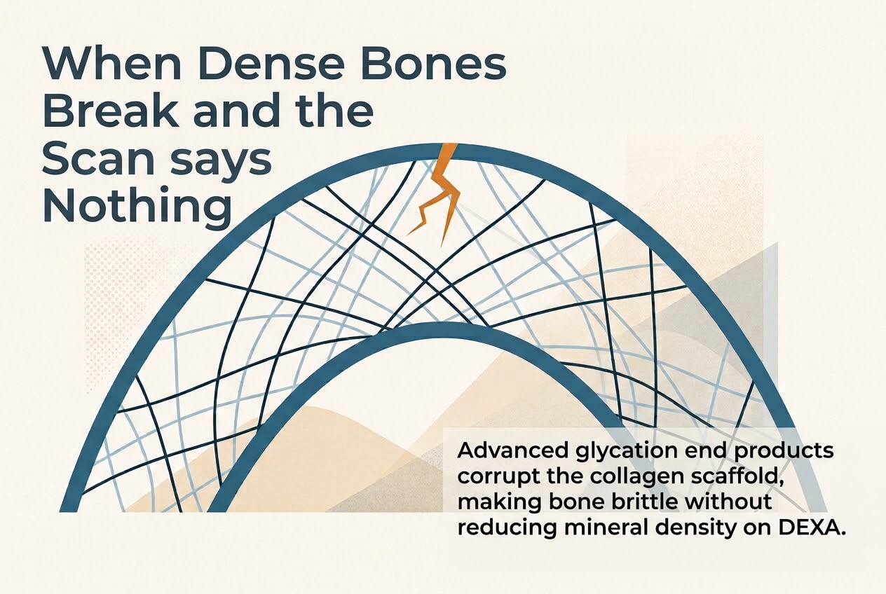
The most extensively studied AGE crosslink in bone is pentosidine, a fluorescent compound formed between lysine and arginine residues via a pentose sugar intermediate. Pentosidine and related AGE crosslinks introduce rigid, abnormal intermolecular bridges within and between collagen fibrils that fundamentally alter their mechanical behavior. Rather than allowing the controlled inter-fibrillar sliding that normally dissipates mechanical energy, AGE-crosslinked collagen fibres are locked in a stiff, brittle configuration. The consequence is a collagen network that, paradoxically, may appear structurally intact under conventional imaging while being markedly compromised in its ability to withstand real-world loading conditions without fracture initiation [5,7].
Insulin Resistance and Skeletal Metabolism: An Overlooked Connection
The relationship between insulin signaling and bone metabolism is bidirectional and biologically significant. Osteoblasts, the principal bone-forming cells express insulin receptors, and insulin signaling directly promotes osteoblast proliferation, differentiation, and survival. In states of insulin resistance, the anabolic drive on bone formation is attenuated. Reduced osteoblast activity translates to lower rates of bone matrix synthesis and, critically, reduced production of the enzymatic crosslinks needed to stabilize newly deposited collagen [6].
Insulin-like growth factor-1 (IGF-1), whose signaling is closely coupled to insulin receptor activity, is a potent stimulus for osteoblast function and bone matrix mineralization. Insulin resistance is frequently accompanied by relative IGF-1 deficiency or attenuated IGF-1 receptor sensitivity in skeletal tissue, further compromising the bone formation response. The net effect is a subtle but progressive deterioration in the quality of newly synthesized bone matrix, even before the cumulative effects of AGE accumulation become clinically apparent [8].
A particularly elegant feedback loop has also been characterized between bone and glucose homeostasis. Osteocalcin, a protein secreted by osteoblasts and a recognized marker of bone formation acts as an endocrine hormone that stimulates pancreatic beta-cell proliferation and insulin secretion, and enhances peripheral insulin sensitivity. In the setting of insulin resistance and impaired osteoblast function, osteocalcin secretion is reduced, potentially creating a self-reinforcing cycle in which metabolic dysfunction worsens bone quality, which in turn further impairs glucose homeostasis. This bidirectionality elevates bone health to the status of a metabolic concern as much as a structural one [8].
Chronic hyperglycaemia, the metabolic hallmark of poorly controlled insulin resistance, adds a further insult through the direct glycation of bone matrix proteins, independent of changes in bone cell activity. It is this chemical modification of existing collagen that may represent the most mechanically consequential consequence of sustained glucose excess [9].
Non-Enzymatic Glycation and the Formation of Advanced Glycation End Products in Bone
The process of non-enzymatic glycation in bone follows the same Maillard reaction chemistry that occurs elsewhere in the body, but bone tissue is uniquely vulnerable due to the exceptionally long half-life of its extracellular matrix proteins. While collagen in soft tissues may be renewed over weeks or months, cortical bone collagen can persist for decades. This extended residence time creates an enormous window of opportunity for cumulative glucose-mediated modification [5].
Glucose and other reducing sugars react with the epsilon-amino groups of lysine residues on collagen molecules through a series of progressive reactions. Early-stage reversible products (Schiff bases and Amadori compounds) give way over time to late-stage irreversible AGEs, including pentosidine, crossline, and carboxymethyl-lysine (CML). Of these, pentosidine has received the greatest scientific attention as a bone quality biomarker because it is measurable in both bone biopsy specimens and in urine, and because its bone concentrations correlate with tissue-level mechanical properties [7,10].
At the biomechanical level, the consequences of elevated pentosidine and related AGE crosslinks are well-documented in human and animal bone specimens. Studies have demonstrated that high pentosidine concentration is associated with reduced post-yield deformation, reduced energy-to-fracture, and decreased toughness, the latter referring to the total energy a material can absorb before fracturing. These are precisely the material properties that protect bone during dynamic loading, stumbling, or falls. In practical terms, a bone rich in AGE crosslinks will crack rather than bend; it will fail suddenly rather than deforming plastically to absorb mechanical energy [5,11].
Importantly, this biomechanical deterioration can occur without any reduction in bone mineral density. AGE crosslinks reside within the collagen phase of bone matrix; they do not demineralize the tissue or reduce its calcium content. A DEXA scan, which measures the attenuation of X-rays by mineral mass, will detect no abnormality in a bone whose collagen is saturated with pathological crosslinks. The tissue will appear dense and will be demonstrably fragile [10].
Animal studies using streptozotocin-induced diabetic rodent models have corroborated these findings in a controlled setting. Diabetic animals consistently exhibit elevated bone pentosidine content, reduced bone toughness, and higher fracture incidence at normal or elevated BMD compared to controls, a direct recapitulation of the human epidemiological paradox [9].
The Paradox of High Bone Mineral Density and Elevated Fracture Risk in Type 2 Diabetes
The epidemiological evidence for a dissociation between BMD and fracture risk in T2DM is now robust and consistent. In a landmark analysis from the Rotterdam Study, Oei et al. examined the relationship between glycaemic control, BMD, and fracture incidence in a large population-based cohort. Patients with T2DM demonstrated BMD values 1.1–5.6% higher than non-diabetic controls at the femoral neck, total hip, and lumbar spine sites typically used in osteoporosis screening. Yet the same patients carried a 47–62% higher risk of non-vertebral fracture. The direction of this dissociation, higher density, more fractures is precisely the opposite of what the conventional density-centric model would predict [3].
These findings are not isolated. A meta-analysis by Vestergaard, encompassing studies of both type 1 and type 2 diabetes, confirmed that patients with T2DM carry a substantially elevated risk of hip and non-vertebral fractures despite having BMD values that are on average higher than non-diabetic reference populations. Schwartz and colleagues similarly documented increased fracture rates in older women with diabetes even after controlling for BMD, body weight (which is generally higher in T2DM and independently correlates with BMD), and other conventional risk factors [2,7].
Several mechanisms contribute to this paradox. First, as detailed above, the accumulation of AGE crosslinks degrades collagen toughness without reducing mineral density. Second, insulin resistance impairs osteoblast-mediated bone matrix quality, independent of bone mass. Third, advanced T2DM is frequently complicated by peripheral neuropathy and orthostatic hypotension, both of which elevate fall risk independent of intrinsic bone fragility. Fourth, cortical bone porosity, detectable by high-resolution peripheral quantitative computed tomography (HR-pQCT) but not DEXA, is increased in T2DM and contributes to structural weakness at cortical sites [5,9,12].
The clinical implication is direct and sobering. A patient with T2DM who presents with a normal or “good” DEXA T-score may have been provided a false reassurance about their fracture risk. The current FRAX algorithm, which incorporates BMD alongside clinical risk factors, does not adequately account for the AGE-mediated deterioration of bone quality and may therefore systematically underestimate fracture probability in this population [3,13].
Limitations of Dual-Energy X-ray Absorptiometry in Assessing Bone Quality
DEXA remains the clinical gold standard for fracture risk assessment by virtue of its wide availability, low radiation dose, and strong population-level association with fracture incidence. Its limitations, however, are fundamental and not merely technical. DEXA measures areal BMD, the projected two-dimensional mineral mass within a defined region of interest and cannot distinguish between cortical and trabecular compartments, assess microarchitecture, or evaluate the properties of the collagen matrix [14].
Bone quality is a multidimensional construct encompassing: (1) microarchitecture, including trabecular number, thickness, spacing, and connectivity; (2) mineral crystal size, degree of crystallinity, and mineralization heterogeneity; (3) collagen fibril organization and the nature of intermolecular crosslinks; and (4) the accumulation of microdamage. None of these dimensions are captured by DEXA. A bone scoring “normal” on DEXA may be architecturally compromised, poorly mineralized, AGE-crosslinked, or riddled with microcracks, all of which reduce fracture resistance independently of mineral mass [11].
It is worth noting that the very pathophysiology of T2DM can produce a DEXA result that is actively misleading. Adipose tissue, which is increased in obese or overweight individuals with T2DM, partially attenuates X-ray beams in a manner that can artificially inflate apparent BMD at measured sites. Concurrently, the hyperinsulinaemic state early in T2DM may temporarily stimulate osteoblast activity and mineralization, contributing to genuinely higher mineral density even as collagen quality deteriorates. The result is a DEXA report that not only fails to detect the true problem, but may actively point in the wrong direction [6,13].
Emerging Assessment Tools and Biomarkers of Bone Quality
Recognition of DEXA’s limitations has stimulated interest in complementary methods that capture dimensions of bone quality beyond mineral density. Several approaches show clinical and research promise.
Trabecular Bone Score (TBS)
TBS is a textural index derived from the pixel grey-level variations within lumbar spine DEXA images. It provides an indirect measure of trabecular microarchitecture without additional radiation exposure or equipment. TBS has been shown to be independently associated with fracture risk in T2DM, adding predictive information beyond BMD alone. Its limitation is that it remains a derived index with indirect mechanistic significance rather than a direct measure of collagen quality or AGE accumulation [15].
High-Resolution peripheral Quantitative CT (HR-pQCT)
HR-pQCT allows three-dimensional, in vivo assessment of cortical and trabecular bone microarchitecture at the distal radius and tibia with spatial resolution sufficient to visualize individual trabeculae. Studies using HR-pQCT have demonstrated that T2DM patients exhibit higher cortical porosity and altered trabecular morphology relative to controls, even at equivalent BMD. This technique is increasingly regarded as the reference standard for research-level bone microarchitecture assessment, though its clinical availability remains limited [9].
Serum and Urinary Pentosidine
Pentosidine, the best-characterized AGE crosslink in bone, is measurable in both serum and urine and correlates with bone tissue pentosidine content in several studies. Elevated urinary pentosidine has been associated with increased fracture risk independently of BMD in older adults. While not yet in routine clinical use, serum or urinary pentosidine measurement may represent a low-invasive method of bone quality surveillance in high-risk individuals [5,10].
Skin Autofluorescence as Surrogate for Tissue AGEs
Because AGEs accumulate in skin collagen as well as bone collagen, skin autofluorescence (SAF), measured by a non-invasive fluorescence spectroscopy device has been proposed as a surrogate marker of systemic AGE burden. SAF has been associated with cardiovascular outcomes in T2DM and is under investigation as a skeletal risk marker. Its utility as a bone-specific tool requires further validation, but it represents an attractive, rapid, and non-invasive screening modality [16].
Clinical and Preventive Implications: Protecting Bone Quality in Insulin Resistance
Understanding insulin resistance as a mechanism of bone fragility shifts the clinical conversation from passive monitoring of BMD to active metabolic management aimed at preserving collagen integrity. Several evidence-informed strategies merit consideration.
Glycaemic Optimization
Reducing chronic glucose exposure is the most direct strategy to attenuate non-enzymatic glycation of bone collagen. Data from the Rotterdam Study indicated that HbA1c was inversely associated with bone quality outcomes: poorer glycaemic control predicted higher fracture risk independently of BMD. A clinically meaningful target is to maintain HbA1c within individualized goals, while avoiding iatrogenic hypoglycaemia, which carries its own skeletal (fall-related) and cardiovascular risks [3].
Pharmacological Considerations in T2DM Management
Not all antidiabetic agents carry equivalent skeletal effects. Thiazolidinediones (TZDs) such as rosiglitazone and pioglitazone, while effective insulin sensitizers, activate PPARγ in bone marrow stromal cells, diverting differentiation away from osteoblasts toward adipocytes and resulting in net bone loss. Metformin, by contrast, activates AMP-activated protein kinase (AMPK) and has been associated with favorable effects on osteoblast differentiation and collagen synthesis in preclinical models. GLP-1 receptor agonists may also confer skeletal benefits via direct osteoblast receptor signaling, though human fracture outcome data remain limited. Clinicians managing T2DM should consider skeletal implications when selecting pharmacological agents [14,17].
Physical Activity and Mechanical Loading
Exercise exerts dual benefits in this context. Mechanically, weight-bearing and resistance exercise stimulate osteoblast activity and bone matrix synthesis via mechanosensory pathways, promoting production of higher-quality collagen with appropriate enzymatic crosslinks. Metabolically, regular physical activity improves insulin sensitivity, reduces chronic glucose exposure, and attenuates AGE accumulation rates. The combination of strength training and aerobic conditioning is therefore a biologically rational and evidence-supported intervention for individuals at elevated skeletal-metabolic risk [12].
Nutritional Strategies
Dietary patterns that reduce postprandial glucose excursions, including reduced refined carbohydrate intake, lower glycaemic index food choices, and adequate dietary fibre can meaningfully attenuate non-enzymatic glycation rates. Protein adequacy is also relevant: collagen synthesis requires sufficient amino acid substrate, and protein insufficiency compounds the effects of reduced osteoblast activity. Emerging evidence supports the potential utility of polyphenol-rich foods and certain B-vitamins (particularly B6, B12, and folate) in modulating AGE formation, though clinical fracture outcome data are not yet available [16].
Anti-Resorptive and Anabolic Therapies
Conventional osteoporosis pharmacotherapy such as bisphosphonates, denosumab, and teriparatide has demonstrated anti-fracture efficacy in general populations. However, their specific utility in T2DM-related bone fragility, where the primary defect is in collagen quality rather than bone mass, requires further investigation. Current evidence suggests that fracture risk reduction may be less predictable in T2DM populations, and treatment decisions should be informed by comprehensive risk assessment beyond BMD alone [1].
Conclusion
The predominant clinical narrative around bone fragility, centered on calcium supplementation and BMD monitoring is scientifically incomplete. For the growing population of individuals with insulin resistance and type 2 diabetes, the principal threat to skeletal integrity is not calcium deficiency or bone thinning, but a progressive biochemical corruption of the collagen scaffold through non-enzymatic glycation and AGE crosslink accumulation. This process renders bone brittle without reducing its mineral density, creating a fracture risk that is invisible to conventional DEXA screening.
The evidence from the Rotterdam Study and multiple corroborating sources is unambiguous: in T2DM, higher BMD and higher fracture risk can and do coexist. This paradox is not an anomaly to be explained away; it is a direct consequence of biology that current clinical frameworks fail to capture. A DEXA scan that reports normal bone density in a person with poorly controlled insulin resistance may be providing meaningless or worse, falsely reassuring information about actual fracture risk [2,3].
Addressing this gap requires a reorientation at multiple levels. Clinically, fracture risk assessment in metabolically compromised individuals should incorporate markers of glycaemic control, emerging tools for bone quality assessment (TBS, HR-pQCT, serum pentosidine), and a comprehensive evaluation of fall risk. Therapeutically, the management of insulin resistance and hyperglycaemia should be recognized as a bone protection strategy, not merely a cardiovascular and renal one. And at the level of patient education, the message must shift: bones are protected not by calcium alone, but by the metabolic health of the matrix in which that calcium resides.
Insulin resistance is eroding bone quality in a growing proportion of the global population. The science now exists to understand why. The clinical and public health challenge is to translate that understanding into action before the fracture occurs, not merely to measure the density of the bone that breaks.
Reference
1. Kanis JA, Cooper C, Rizzoli R, Reginster JY. European guidance for the diagnosis and management of osteoporosis in postmenopausal women. Osteoporos Int. 2019;30(1):3–44. doi:10.1007/s00198-018-4704-5
2. Vestergaard P. Discrepancies in bone mineral density and fracture risk in patients with type 1 and type 2 diabetes—a meta-analysis. Osteoporos Int. 2007;18(4):427–44. doi:10.1007/s00198-006-0253-4
3. Oei L, Zillikens MC, Dehghan A, Buitendijk GH, Castaño-Betancourt MC, Estrada K, et al. High bone mineral density and fracture risk in type 2 diabetes as skeletal complications of inadequate glucose control: the Rotterdam Study. Diabetes Care. 2013;36(6):1619–28. doi:10.2337/dc12-1achiev (PMID: 23315602)
4. Viguet-Carrin S, Garnero P, Delmas PD. The role of collagen in bone strength. Osteoporos Int. 2006;17(3):319–36. doi:10.1007/s00198-005-2035-9
5. Saito M, Marumo K. Collagen cross-links as a determinant of bone quality: a possible explanation for bone fragility in aging, osteoporosis, and diabetes mellitus. Osteoporos Int. 2010;21(2):195–214. doi:10.1007/s00198-009-1066-z
6. Schwartz AV. Diabetes mellitus: does it affect bone? Calcif Tissue Int. 2003;73(6):515–9. doi:10.1007/s00223-003-0023-7
7. Schwartz AV, Sellmeyer DE, Ensrud KE, Cauley JA, Tabor HK, Schreiner PJ, et al. Older women with diabetes have an increased risk of fracture: a prospective study. J Clin Endocrinol Metab. 2001;86(1):32–8. doi:10.1210/jcem.86.1.7139
8. Saito M, Kida Y, Kato S, Marumo K. Diabetes, collagen, and bone quality. Curr Osteoporos Rep. 2014;12(2):181–8. doi:10.1007/s11914-014-0202-5
9. Burghardt AJ, Issever AS, Schwartz AV, Davis KA, Masharani U, Majumdar S, et al. High-resolution peripheral quantitative computed tomographic imaging of cortical and trabecular bone microarchitecture in patients with type 2 diabetes mellitus. J Clin Endocrinol Metab. 2010;95(11):5045–55. doi:10.1210/jc.2010-0994
10. Yamamoto M, Yamaguchi T. Mechanisms of increased bone fragility in diabetes. Endocr J. 2010;57(10):843–53. doi:10.1507/endocrj.K10E-220
11. Farr JN, Khosla S. Determinants of bone strength and quality in diabetes mellitus in humans. Bone. 2016;82:28–34. doi:10.1016/j.bone.2015.07.027
12. Wongdee K, Charoenphandhu N. Osteoporosis in diabetes mellitus: possible cellular and molecular mechanisms. World J Diabetes. 2011;2(3):41–8. doi:10.4239/wjd.v2.i3.41
13. Hough FS, Pierroz DD, Cooper C, Bonjour JP; IOF CSA Bone and Diabetes Working Group. MECHANISMS IN ENDOCRINOLOGY: Mechanisms and evaluation of bone fragility in type 1 diabetes mellitus. Eur J Endocrinol. 2016;174(4):R127–38. doi:10.1530/EJE-15-0973
14. Lecka-Czernik B. Bone loss in diabetes: use of antidiabetic thiazolidinediones and secondary osteoporosis. Curr Osteoporos Rep. 2010;8(4):178–84. doi:10.1007/s11914-010-0027-y
15. Garnero P. New developments in biological markers of bone metabolism in osteoporosis. Bone. 2014;66:46–55. doi:10.1016/j.bone.2014.05.016
16. Brownlee M. Biochemistry and molecular cell biology of diabetic complications. Nature. 2001;414(6865):813–20. doi:10.1038/414813a
17. McCarthy AD, Cortizo AM, Sedlinsky C. Metformin revisited: does this regulator of AMP-activated protein kinase secondarily affect bone metabolism and prevent diabetic osteopathy? World J Diabetes. 2016;7(6):122–33. doi:10.4239/wjd.v7.i6.122